Describe the Use of Dental Imaging
Dental radiographs normally expose the individual patient to relatively low doses of ionising radiation but unfortunately dental radiographs are used much more frequently than medical radiographs and tend to be used on a generally younger population of patients who are more at risk of harm from ionising radiation. In comparison to CBCT a similar image quality with complementary contrast was.

Types Of Dental Radiographs And Their Uses Dentalnotebook
The application of computer imaging software used in dentistry has become one of the main tools for dentists in providing superior services to their.
. 3D Imaging Using CBCT. Start studying the chapter 38 dental flashcards containing study terms like The uses of dental images include the detection of exposure to radiation the x-ray was discovered in 1895 by and more. Orthodontists and dentists use Cone Beam Computerized Tomography CBCT which evolved from CAT.
Willis July 21 2017 Short- Answer Questions 1. Evaluate growth and development. In 1973 computed tomography CT created images by combining x-ray and computer technology to capture thin slices of tissue1 After that magnetic resonance imaging MRI allowed soft tissue analysis.
Provide information during dental procedures such as root canal therapy Document a patients condition at a specific time. Caries commonly known as tooth decay that can develop between your teeth or under fillings Diseases in your jaw bones Gum disease also known as periodontal disease Infections under your gums Some types of tumors. Near-infrared imaging offers a non-ionizing alternative for dental analysis.
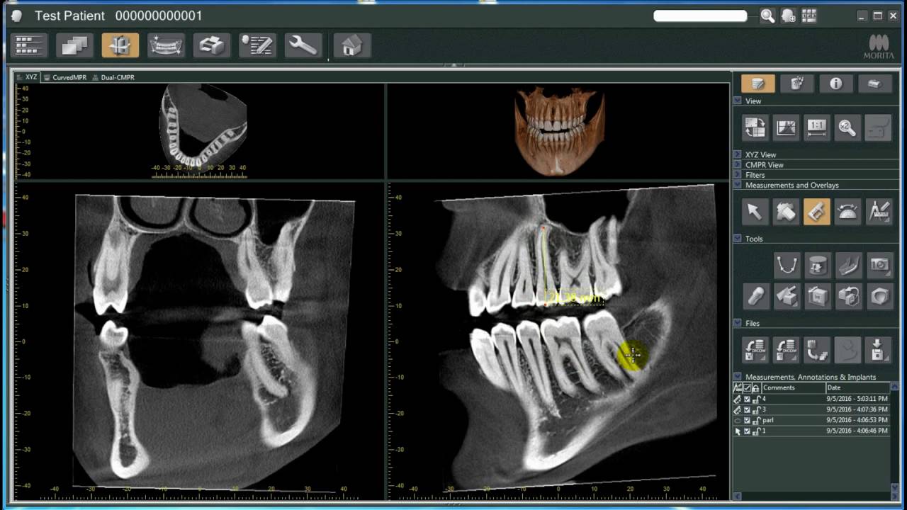
Also many dental offices are now using digital images instead of film. The cone beam computed tomography is used to view the area of the head and neck in three dimensions and it is used to find the exact placement of implants the buccallingual position of impacted teeth to be removed and determination of the exact location of the mandibular nerve before surgery is done. To find out how you can benefit from 3D images make an appointment by calling our office at 951 244-9495 or going online today.
Place and secure the. Dental X-rays or radiographs are images that are captured of your teeth and subsequently used by your dentist for assessing your oral health. Memorize flashcards and build a practice test to quiz yourself before your exam.
Digital radiography is a type of X-ray imaging that uses digital X-ray sensors to replace traditional photographic X-ray film producing enhanced computer images of teeth gums and other oral structures and conditions. Katherine Moran Radiology Mrs. We describe the construction and.
X-ray imaging including dental CBCT provides a fast non-invasive way of answering a number of clinical questions. Image receptor must be placed away from the teeth and toward the middle of the mouth Explain why an image receptor holder is necessary with the paralleling technique. Intraoral digital imaging has evolved from an experimental and sometimes disparaged technique in the mid 1980s to a reliable and ubiquitously used technology today.
Two-dimensional radiographs are commonly used for evaluating sub-surface hard structures of teeth but they have low sensitivity for early caries lesions particularly those on tooth occlusal surfaces and they are also frequently refused by patients over safety concerns. Hauser uses 3D scans to diagnose a wide variety of dental problems. In the case of 3D dental imaging the advantages are clear granting practitioners and patients alike a better clinical experience.
Elements of effective imaging. Additionally the scans are used in conjunction with computer aided design CAD programs and CEREC to craft precise dental restorations. Using dental radiography your dentist can detect.
Uses of digital imaging-To detect lesions diseases and conditions of teeth and surrounding structures-Confirm or classify suspected disease-Provide information during dental procedures root canal therapy instrumentation and surgical placement of. The uses of dental imaging are checking patients oral health and making it clearer to diagnose certain teeth problems such. Dental CBCT images provide three-dimensional 3-D information rather than the.
Describe the purpose and uses of cone beam computed tomography. Not only is time saved in the development process reducing the amount of radiation by as much as 80. Detect dental caries in the early stages.
An offshoot of dental X-rays is 3D imaging. Describe the use of dental imaging. The 3 Most Common Types of Dental X-Rays There are several different types of x-rays that your dentist in Buckhead may use to look at various parts of the mouths anatomy.
One of our more frequent requests is for more information on dental imaging why we use them different types and especially their safety. A 3D scan can help gain a better view of bone structures such as adjacent root positions in order to locate canals and root fractures as well as provide the ability to more. Identify bone loss in the early stages.
There are many advantages for use of digital radiographic techniques in dentistry one of the chief ones being patient dose reduction. Dental professionals today are increasingly using digital dental radiographs digital X-rays to better detect diagnose treat and monitor oral conditions and diseases. The patients head must be positioned so that the upper arch is parallel to the floor and the midsagittal midline plane is perpendicular to the floor.
Compared to X-ray imaging however so far the spatial resolution of MRI is lower and the scan time is longer. Describe how to prepare a patient for dental imaging. Karen Munoz Jasmine Angel Procedural Steps Adjust the hardest to support and position the patients head.
In this contribution we describe wireless inductively-coupled intraoral coils whose local sensitivity enables high resolution MRI of dental soft tissue. Describe the uses of dental imaging. Locate abnormalities in surrounding hard and soft tissues.
A dental 3D scan allows clinicians to view dental anatomy from different angles. Dentists are using dental imaging to perform dental services that were unimaginable a few years ago. On the word of the best dentist in India dental X-rays are employed with low radiation levels for showing images of areas constituting the inners of your teeth and gums.
The history of dental imaging began in the late 1800s with the development of the x-ray image. Imaging technology aids in the diagnosis of endodontic pathosis and canal morphology assessing root and alveolar fractures in the analysis of resorptive lesions identification of the pathosis of non-endodontic origin and. To generate images that can be used in the diagnosis and assessment of dental disease-detect lesions diseases conditions of teeth and surrounding structures-confirm or classify suspected disease-provide info during dental procedures root canal therapy instrumentation and surgical placement of implants-to evaluate growth and development.

Types Of Dental Radiographs And Their Uses Dentalnotebook
What Is 3d Dental Imaging North Brand Dental Glendale Ca 818 244 7215

Situs Inversus Short Form Of The Latin Situs Inversus Viscerum Is A Term Used To Describe The Radiology Imaging Human Anatomy And Physiology Medical Dental


Comments
Post a Comment